Tuesday, April 28, 2009

"Dissection: Photographs of a Rite of Passage in American Medicine 1880–1930," Blast Books, 2009


I just got my hands on the new Blast Books offering Dissection: Photographs of a Rite of Passage in American Medicine 1880–1930, by John Harley Warner and James M. Edmonson. You might be familiar with Blast Books from other wonderful publications such as the Mütter Museum calendars and books, and this book is just as memorable and essential as those previous releases. The book is large-scale, beautifully produced, and filled with fascinating and haunting photographs (such as those shown above; click on images see larger versions) so wonderfully reproduced that you feel you could walk right into them. The introductory essays by Warner and Edmonson do an admirable job of parsing this overlooked genre of photography and providing a sense of context for images so unusual and sensational to the modern eye.

I asked Laura Lindgren, co-publisher of Blast Books and the editor and designer of this tome, to give me some information about the book and send me a few of her favorite images (which I have featured above). Here's the press release she sent:
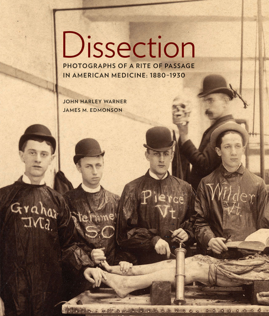
DISSECTION Photographs of a Rite of Passage in American Medicine 1880–1930
by John Harley Warner and James M. Edmonson
Blast Books, 2009

Featuring 138 rare, historic photographs, Dissection is a “landmark book” (Ruth Richardson) that reveals a startling piece of American history, the rite of passage into the mysteries of medicine captured in photography.

From the advent of photography in the 19th century and into the 20th century, medical students, often in secrecy, took photographs of themselves with the cadavers that they dissected: their first patients. The photographs were made in a variety of forms, from proud class portraits to staged dark-humor scenes, from personal documentation to images reproduced on postcards sent in the mail. Poignant, strange, disturbing, and humorous, they are all compelling.

These photographs were made at a time when Victorian societal taboos against intimate knowledge of the human body were uneasily set aside for medical students in pursuit of knowledge that could be gained only in the dissecting room. Dissection, writes Mary Roach, “documents—in archival photographs and informed, approachable prose—a heretofore almost entirely unknown genre, the dissection photograph.”

“Without looking,” writes John Harley Warner, “we cannot see an uncomfortable past and begin to understand the legacies that American doctors and patients live with today.” That uncomfortable past saw the gradual passing of state laws, from 1831 to 1947, to govern the awkward business of cadaver supply—ever inadequate—bringing an end to reliance on professional “resurrectionists,” grave robbing, and dissection as an extended punishment for murder and as a consequence of poverty.

As James Edmonson notes, “Unsettling though these images may be, they are a thread connecting us to the shared experience among medical professionals over generations. . . . As medical schools explore alternatives to human dissection, this rite of passage may disappear.”

Together, the remarkable archival photographs and illuminating essays in Dissection present the astonishing social realities of the pursuit of medical knowledge in 19th- and early-20th-century America.

About the authors: John Harley Warner, PhD, is an historian who focuses chiefly on American medicine and science. In 1986 he joined the Yale faculty with a primary appointment in the School of Medicine, where he is now Avalon Chair of the Section of the History of Medicine. James M. Edmonson, PhD, is Chief Curator of the Dittrick Medical History Center and Museum of Case Western Reserve University in Cleveland.

About Blast Books: Celebrating our 20th anniversary in 2009, Blast publishes books on cultural and historical phenomenons, from Mütter Museum to Mental Hygiene. Visit www.blastbooks.com.

Praise for Dissection
“An extraordinary collection of photographs that makes even today’s flesh-and-blood anatomy laboratories look tame. . . . Forget the truckloads of grandiose prose that has been spun about the art and science of medicine over the centuries: one look at this picture [page 188] and you understand what it is all supposed to be about.”—Abigail Zuger, MD, The New York Times

“This is the most extraordinary book I have ever seen [and] the perfect coffee table book for all the households where I’d most like to be invited for coffee.”
—Mary Roach, author of Stiff and Bonk

“A truly unique and important book [that] documents a period in medical education in a way that is matched by no other existing contribution.”
—Sherwin Nuland, MD, author of How We Die

“What a spectacular book! Warner and Edmonson have amassed an extraordinary array of old dissecting-room photographs. Together, the authors have created a landmark book in the history of dissection in America.”
—Ruth Richardson, D.Phil., author of Death, Dissection and the Destitute and The Making of Mr. Gray’s Anatomy
The book has been (rightfully) been getting a lot of excellent press; see the New York Times review by clicking here and the Slate.com writeup by clicking here; You can also read Bioephemera's thoughtful response here. It is nice to see that audiences are responding so positively and with great interest to what, I was afraid, might be seen as rather off-putting images to the larger public.

Also, I just got back from the MeMa (Medical Museum Association) meeting at the Dittrick Medical History Center in Cleveland, where, after giving a presentation on how medical museums can utilize the web to reach new audiences, I was fortunate enough to see some of the original photographs on display in an exhibition called "Haunting Images: Photography, Dissection, and Medical Students." The Dittrick has created an online version of the exhibition which you can access by clicking here.

I simply cannot recommend this book highly enough. It is absorbing, transporting, beautiful, and thought-provoking. I have never seen another collection like it. You can order a copy of the book (at the moment only about $32 with amazon's discount!) by clicking here. You can see Blast Books other medically oriented illustrated books by clicking here.

Images, top to bottom (after cover):
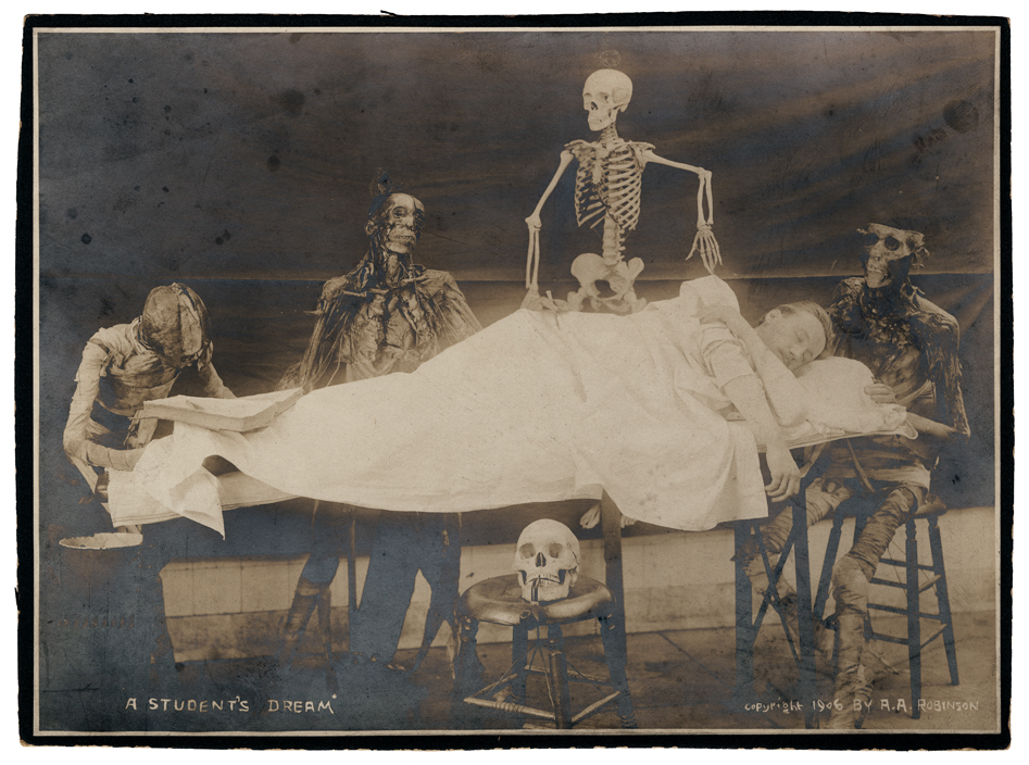
1. “A Student’s Dream,” 1906. Copyright marked A. A. Robinson. A standing skeleton joins seated cadavers, preparing to dissect a sleeping medical student. Iconographic elements that by 1906 had become common-place in dissecting room group portraiture are gathered together in this scene: the book propped open on the “subject’s” feet, the skull resting on a stool in front of the table, and a pipe fitted between the skull’s teeth. DMHC. Used by permission of Blast Books, Inc.

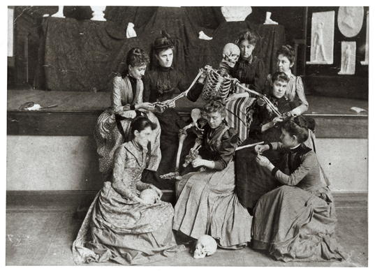
2. Western Female Seminary (renamed the Western College for Women in 1904), Oxford, Ohio. Once removed from dissection, the students at this daughter school of Mount Holyoke studied hygiene, anatomy, and physiology with the use of a skeleton, manikin, and the plaster casts that appear behind the tableau. The instructor probably was Robert L. Rea, M.D., who was resident at the women’s college and later became professor of anatomy at Rush Medical College in Chicago. Miami University Libraries, Western College Memorial Archives, Miami University, Oxford, Ohio. Used by permission of Blast Books, Inc.

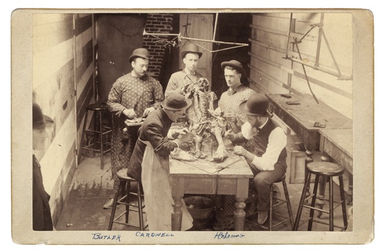
3. Probably Rush Medical College, Chicago, ca. 1915. Private collection. Used by permission of Blast Books, Inc.

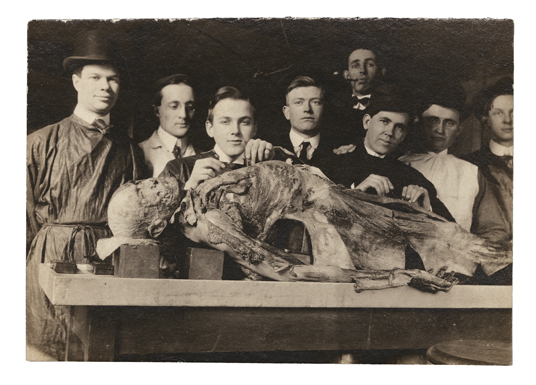
4. Cabinet card dated February 9, 1887, Willamette University Medical Department, Portland, Oregon. A man (wearing a bowler hat, like the dissectors) looks on from the lower left of this cramped space. The medical school had just moved from Salem, Oregon, to Portland. The photograph belonged to Otis D. Butler, age twenty-five when the photograph was taken, who died in 1926 as a result of an infection of the hand received during a surgical operation. DMHC. Used by permission of Blast Books, Inc.

Monday, April 27, 2009

The Agricultural Museum, Cairo, Egypt, Est. 1930


Cairo based Morbid Anatomy Reader Oliver Wilkins recently sent me an email telling me about--and linking to photos of--one of his favorite museums, the amazing looking Agricultural Museum of Cairo, Egypt. I asked him to tell me a bit more about the museum, which I had never heard of; Here is what he had to say:
The Cairo Agricultural Museum was founded in 1930 in the palace of Princess Fatima, in what is now Dokki. With it's numerous buildings, laboratories, themed halls, botanical gardens, cinema and greenhouses, it must have been quite a complex. Little has changed since it was established including the entrance fee of 10 piastres (around one cent).

The museum has fallen into disrepair and many sections are now closed, although the caretaker will take you behind the scenes for a small tip. Highlights include the bread museum, a room of wax models of typical meals of Egypt, a large selection of preserved animals, digestion displays including an inflated cows stomach, and the museum of disease and health. Most of the exhibits date from the 1930s and include exquisite wax models, hand drawn posters and curious taxonomy.

Lack of investment and minimum wages have not helped. A small tip is demanded to see the stuffed Lion, his legs broken from being dragged across the floor and various exhibits are damaged or missing. The Mathaf Al-Zira'ee is a museum that should be in a museum, who knows how long it will be until it goes the way of Cairo's other lost museums? See it while you can.
Click here to find out more about the museum. Click here to see Oliver's complete photoset (these are just a small few of what you will find there; many more amazing photos to be found there.)

Sunday, April 26, 2009

Images from the History of Medicine on Bibliodyssey


Here are some highlights from a recent Bibliodyssey post entitled "Images from the History of Medicine;" you can view the full post (with many more images) by clicking here. All images drawn from the National Library of Medicine's image database, which you can visit by clicking here.

Images, top to bottom: 1. Signs of Character, Drawn and Published by R. Degranza Pease M.D. 1843. Phrenology poster which shows a profile with labeled sections in an elaborate border. 2. The Anatomist by Leonard Baskin (20th cent.), Half-length figure of a man holding a model of a skeleton; partial anatomical chart to the right. 3. The Human Body and the Library as Sources of Knowledge, Fronispiece in 'Tabulae Anatomicae' by Johann Adam Kulmus (early 18th cent.) Interior view of a library with allegorical figures; a body rests on a dissections table in center; a skeleton stands in an alcove to the right; surgical instruments are arranged on a pedestal in the foreground; bookshelves fill the background.

Wednesday, April 22, 2009

More Wonderful Images from the National Museum of Health & Medicine's Flickr Page


Here are three more wonderful images (click here to view others featured in recent posts) from the National Museum of Health & Medicine's Flickr collection; the top two images have captions that read: "Drawing: anatomical charts. [Possibly World War 2 era.]" the ottom image: "Unidentified operation (tying off the carotid artery?) during World War I. Artwork by Summers. Selected by Mike."

To see more images from their collection, visit their astounding Flickr collection by clicking here. You might also want to check out their blog A Repository for Bottled Monsters.

Tuesday, April 21, 2009

Second Nature: International Journal of Creative Media, Vol 1, No 1 (2009)


Shirlee Saul, proprietress of Memory Palace and Curiosity Cabinet (as well as a really wonderful Flickr photostream) is now also the editor of a new journal "Second Nature: International Journal of Creative Media." Volume One, Issue One--which has just launched, in web and PoD Lulu format--is themed "Role Models," and includes a beautifully presented piece on Anatomical Theatre, a photographic exhibition of medical museums that I launched a few years ago. The rest of the journal looks to be pretty interesting as well; here is a description of the journal and this particular issue, from the Lulu page:
Second Nature: The International Journal of Creative Media is a new open access, peer-reviewed online journal that explores the distinctive particulars of and interconnections between textual, visual, aural and interactive creative research and practices. This issue, 'Role Models', is headlined by a keynote article 'Down with the Design Professions!' from eminent MIT scholar, Prof. William J. Mitchell. Articles by other scholars and researchers tease out some of the issues and ideas suggested by Mitchell, interrogating the theme 'role models' from a variety of angles. The journal's 'Projects' include suites of photography -- suggesting non-linguistic approaches to the conceptual and creative challenges and opportunities that the past poses for the present.
You can peruse the entire journal online by clicking here. You can view the Shirlee's piece on Anatomical Theatre by clicking here; click here for more on the exhibition. You can order a hard copy of the journal--full color and 200 pages!--by clicking here. The theme of issue 2, incidentally, will be "Mobile Gaming and Haptic Screen Cultures;" if you are interested in submitting a paper or in finding out more, click here.

Monday, April 20, 2009

Pathological Waxworks, "Palacio de la Escuela de Medicina", Mexico City


Morbid Anatomy reader Jason Plumb just alerted me to an amazing photoset (from which the above images are drawn) of pathological waxes on display at the Palacio de la Escuela de Medicina in Mexico City. The photos were taken by Daniel Menche, who apparently was commissioned to give a performance (!!!) in the museum!

You can read more about Menchel's experience on his blog by clicking here; you can see the whole photoset (which I highly recommend you do! This is just the tip of the proverbial iceberg!) by clicking here. You can visit the museum website by clicking here.

Thanks, Jason, for sending this along!

Saturday, April 18, 2009

Hypochondria, "Villette," Charlotte Brontë, 1853


There sat a silent sufferer — a nervous, melancholy man. Those eyes had looked on the visits of a certain ghost — had long waited the comings and goings of that strangest spectre, Hypochondria. Perhaps he saw her now on that stage, over against him, amidst all that brilliant throng. Hypochondria has that wont, to rise in the midst of thousands — dark as Doom, pale as Malady, and well nigh strong as Death. Her comrade and victim thinks to be happy one moment — " Not so," says she ; "I come." And she freezes the blood in his heart, and beclouds the light in his eye.

Villette, Charlotte Brontë, 1853
Read the full text of Villette (a great read through and through!) by clicking here. You can also purchase a hard-copy by clicking here. And a special thanks to my good friend Catherine for lending me a copy of this book. It was truly a perfect traveling companion.

Image: "The Hypochondriac," by Thomas Rowlandson, 1788. from the online exhibiton "Before Depression, 1660-1800". Click on image to see larger version.

Friday, April 17, 2009

Modern Medical Marvel: Fir Grows in Mans Lung, 2009, Ural Mountains


In a story that brings to mind early medical hoaxes such as Mary Tofts' miraculous, doctor-verified birthing of a litter of rabbits in 1726, doctors in the Ural Mountains claim to have recently opened up the chest of a man suspected of having lung cancer and finding... a small fir tree.

From the article in the Gaurdian:
The annals of medical anomalies bulge with stories from far-flung places where the idea of a reliable source is a chap sitting on a gate in a goatskin fleece who waves to passersby, even if there are none. And so to the Urals, where medics are reported to have removed a tiny fir tree from a man's lung, after he complained of chest pains. Before doctors opened him up, they were convinced he had lung cancer. Now, they're convinced he inhaled a seed, which sprouted inside him.

Surgeon Vladimir Kamashev at Izhevsk hospital was about to remove a large part of 28-year-old Artyom Sidorkin's lung, when he took a closer look, according to reports. He was stunned to see a 5cm-long spruce inside, the Russian news agency Pravda says.

A spokeswoman for the Royal Botanic Gardens in Kew, London, is flummoxed. "A seed might be able to germinate in the damp, dark conditions of a lung, but it's still bizarre," she says.

The gruesome photo [see bottom image] released with the story claims to show the spruce jutting from a clump of Sidorkin's lung tissue. The plant looks firm and healthy, with bright green needles. It's as if it had been grown in the best soil with plenty of sunlight. It lacks roots in the way fresh clippings do.

Lungs are good at getting rid of unexpected visitors. They are lined with mucus that traps everything from mould spores to flies. This is pushed out of the lungs by tiny hairs called cilia. You end up coughing it out, or swallowing it.

"The closest I've heard to this are balls of mould that grow in patients who have abnormalities in their lungs," says Simon Johnson, a reader in respiratory medicine at Nottingham University. "They can get up to a few centimetres, but you would know because you would be coughing up blood."
Both images are drawn from the Geekologie post "Gross!: Man Grows Small Fir Tree In Lung;" click here to see that post. Read the full Gaurdian story by clicking here. To find out more about Mary Tofts, click here. You can watch a short, unintentionally surrealistic video on the story (after waiting through an annoying commercial) by clicking here.

Thanks to John Troyer, Death and Dying Practices Associate, Centre for Death and Society at the University of Bath) for bringing this story to my attention!

Thursday, April 16, 2009

"Medi-Vaudeville," Ullage Group at Jalopy, Sunday, April 19th


I just found out about a wonderful sounding event taking place in Red Hook, Brooklyn this Sunday, April 19th. I will definitely be going; hope to see you there! Details below:

The Ullage Group offers its fourth afternoon of cultural curiosities, "Medi-Vaudeville." The title refers to the book that Charles Fort planned to write during his final days in the hospital; for, this time around, it's all medical.

Doug Skinner and Dr. Mamie Caton will talk about alternative medicine, evidence-based medicine, and the difficulty of applying scientific methodology to medical procedures. Lisa Hirschfield will present a selection of medically-themed ditties, ranging from 18th century satirical broadside ballads to 20th century patent medicine jingles. Anthony Matt will speak about turn of the century non-invasive medical technologies, which claimed to cure everything from hair loss to cancer by projecting various spectrums of light onto an ailing patient. His talk will explore the medical pursuits of Nikola Tesla, Royal Rife, and Dr. Dinshah Ghadiali. He will also demonstrate a working vintage violet ray generator.

All of this will take place on Sunday, April 19th, at 5 pm, at Jalopy Theatre, 315 Columbia St., Red Hook, Brooklyn. We charge a pittance, $5, to cover our expenses. We would like to point out that you may also hobnob with fellow ullage fanciers, and purchase stimulating beverages; and that the benefit to your overall health and happiness cannot be quantified. And you don't need to make an appointment!

For directions to Jalopy, see www.jalopy.biz.

For more on the Ullage Group, see www.ullagegroup.com.
Image: "The Sailor and the Quack Doctor!!," by George Moutard Woodward, ca.1760-1809, Possibly from Eccentric Excursions, 1796. Found on the wonderful National Library of Medicine website. Click here for full record.

Wednesday, April 15, 2009

"Nature Morte: Formaldehyde Photography and the New Grotesque," Mark Dery, 1999; Free Download!


When I was working on my photographic exhibition Anatomical Theatre, I looked everywhere in an attempt to find writers or critics who discussed artworks inspired by medical museums, anatomical models, and preserved human specimens. In all my searching, I came across only one writer, thanks to my good friend Matt Haber, who sent his book my way; his name is Mark Dery, and you might remember from this recent Morbid Anatomy post.

In his book The Pyrotechnic Insanitarium: American Culture on the Brink, Dery explores this topic in three essays comprising the section "Dead Meat," the most directly medical-museumy of which is the wonderful essay "Nature Morte: Formaldehyde Photography and the New Grotesque." The good news is that Mark just linked from his Shovelware blog to a free, down-loadable PDF of the essay, images included! I highly recommend giving this essay--if not the entire book--a read. The essays are full of fascinating information compellingly related to artists whose work lies in the realm of--in Dery's memorable phrase--the "Pathological Sublime."

More about the essay, from the description on his blog post:
In "Nature Morte: Formaldehyde Photography and the New Grotesque," a chapter from his meditation on the millenial zeitgeist, The Pyrotechnic Insanitarium: American Culture on the Brink (Grove/Atlantic: 1999), cultural critic Mark Dery analyzes the abject aesthetic he calls the New Grotesque, exemplified by the photography of Joel-Peter Witkin and Rosamond Purcell, Nine Inch Nails videos such as "Closer," David Fincher's movie Seven, and most notably the obscure subculture of medical-museum tourists whose mecca is the Mutter Museum in Philadelphia. "If the Enlightenment ushered in the 'disenchantment of the world,' as Max Horkheimer and Theodor Adorno put it, postmodernism returns us to the age of wonder---and terror," writes Dery. "Now, as we return to a world of gods and monsters, there's a burgeoning fascination, on the cultural fringes, with congenital deformities, pathological anatomy, and other curious from the cabinet of wonder."

Drawing on Lawrence Weschler's study of the Museum of Jurassic Technology (Mr. Wilson's Cabinet of Wonder), Gwen Akin and Allan Ludwig's seminal essay "Repulsion: Aesthetics of the Grotesque," Julia Kristeva's notion of the abject, Wolfgang Kayser's landmark study of the grotesque, and Oliver Wendell Holmes's 1845 paean to "worshippers of morbid anatomy," Dery theorizes the Pathological Sublime, an aesthetic emotion that is equal parts horror and wonder, inspired by works of art (or nature) that hold beauty and repulsion in perfect, quivering tension. The Pathological Sublime is the sensation Emily Dickinson had in mind when she wrote, "'Tis so appalling---it exhilarates..."
You can see Dery's original post by clicking here. You can access the PDF directly by clicking here. You can buy a copy of the book by clicking here.

Image: From Anatomical Theatre. Museum of Anatomical Waxes “Luigi Cattezneo” (Museo Delle Cere Anatomiche “Luigi Cattaneo”): Bologna, Italy; Wax model of the eye apparatus and related structures; Clemente Susini, late 18th or early 19th Century

Tuesday, April 14, 2009

Alphonse Bertillon (1853—1914)


If you are a fan of the illustrious history of human classification systems based on physical measurement, you might enjoy E-L-I-S-E's recent collection of visuals related to Alphonse Bertillon (1853—1914), creator of "anthropometry" the mugshot, and the conventional crime scene photograph.

You can visit the original post, from which the above images are drawn, by clicking here. You can find out more about Bertillon by clicking here.

Thursday, April 9, 2009

Blind Pony Presents at Observatory, Good Friday, 7:00 PM


Hi all. Sorry for the recent silence of Morbid Anatomy; I have been at an all-consuming desk job that does not allow me much free time. I would like to break this unintentional silence with an invitation to the next Observatory event, taking place on Good Friday (that's this Friday, FYI...) and organized by co-Observator Herbert Pfostl of Blind Pony. Following is his invitation. Hope to see you there!

Please join me at Observatory
for Blind Pony's first evening:
*

B L I N D P O N Y
P R E S E N T S
*On Good Friday, April 10th at 7 o' clock pm,*
*"T H E D E V I L B R O K E M Y A R M"*
I M A G E S , I N J V R Y , & S O N G
by
P E R F V G I V M.
*Performed and Displayed in the*
OBSERVATORY
543 Union Street (at Nevins) Brooklyn, New York
Entry via Proteus Gowanus Interdisciplinary Gallery and Reading Room;
go through back door of gallery, then take a left to find event.
Directions call 718.243.1572.

http://blindpony.blogspot.com/

Thursday, April 2, 2009

Reminder: Tomorrow Night: "Italian Wax Anatomical Models in European Collections," Lecture at Observatory


Just a friendly reminder: Tomorrow night--Friday, April 3rd--will be our next presentation at Observatory; see following for details. This looks to be a good one; hope you can make it!
Morbid Anatomy presents at Observatory:
"Italian Wax Anatomical Models in European Collections"
Marie Dauenheimer, Trustee of the Vesalius Trust and Medical Illustrator
Tomorrow, Friday, April 3rd
Doors open at 7:00; Presentation at 7:30 PM

This illustrated presentation will examine the art and history of the wax anatomical models of the “Museo Zoologico La Specola” in Florence, Italy. Over 2,000 wax models of human anatomy were created by the museum's “Wax Modeling Workshop” from the mid 18th to early 19th century, and the products of their labor--best known to modern audiences through Tachen's Encyclopaedia Anatomica--
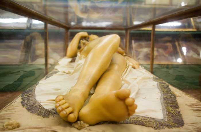
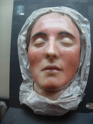
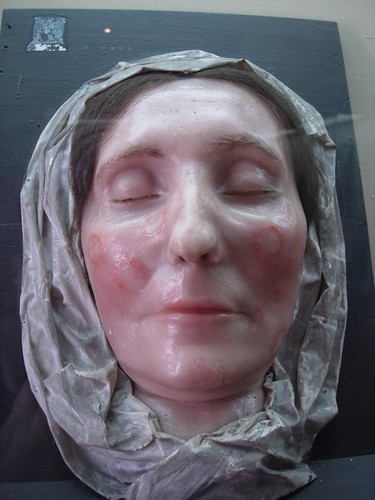
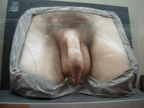
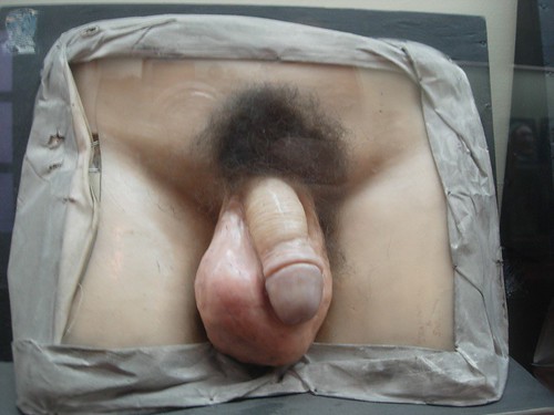
are considered by many to be the finest anatomical waxworks in the world.

This presentation will address how and why these anatomical masterpieces were created, the artists and anatomists who created them, and the place of these collections in the history of anatomical art. The wax anatomical models of Bologna, which pre-date those of “La Specola,” will also discussed.

Marie Dauenheimer is a board-certified Medical Illustrator living in the Washington D.C. area. She is also a trustee on the board of the Vesalius Trust, a non-profit organization which works to support education and research in medical illustration and related visual communication professions. Marie leads the Vesalius Trust Art and Anatomy tours, which are educational tours of important anatomical museums throughout the world. This year's tour--from October 27 to November 8th--will feature museums in Florence (including "La Specola"), Bologna, Venice, and Padua; for more information about this tour, see this recent post. You can see some of Marie's work by clicking here.

Practical Details
"Italian Wax Anatomical Models in European Collections"
Marie Dauenheimer, Trustee of the Vesalius Trust and Medical Illustrator
Friday, April 3rd
Doors open at 7:00; Presentation at 7:30 PM
Admission: Free
Location: Observatory
543 Union Street (at Nevins) Brooklyn, New York 11215
Entry via Proteus Gowanus Interdisciplinary Gallery and Reading Room; go through back door of gallery, then take a left to find event. Directions here or call 718.243.1572.

To learn more about Observatory, click here; you can also visit our under-construction website by clicking here. To get on mailing list, or if you if you might be interested in presenting an event in the future, email me by clicking here.

Image: Wax Anatomical Venus "La Specola;" Part of the exhibition "Anatomical Theatre."