

I am so excited to announce that the amazing Barts Pathology Museum has launched a blog! On the blog, curator Carla Valentine will regularly report on her detailed research into the over 5,000 fascinating specimens she cares for at the Grade II listed Barts Pathology Museum at St Bartholomew’s Hospital, West Smithfield, London.
To celebrate the launch, I asked Carla if she might like to write a guest post about one of my favorite pieces from the collection which I had admired in the recent Museum of London's "Doctors, Dissection and ResurrectionMen" exhibition.
Following is her post; If you like what you read (and see!) I urge you to check out the wonderful--and delightfully image heavy--blog by clicking here.
Following is her post; If you like what you read (and see!) I urge you to check out the wonderful--and delightfully image heavy--blog by clicking here.
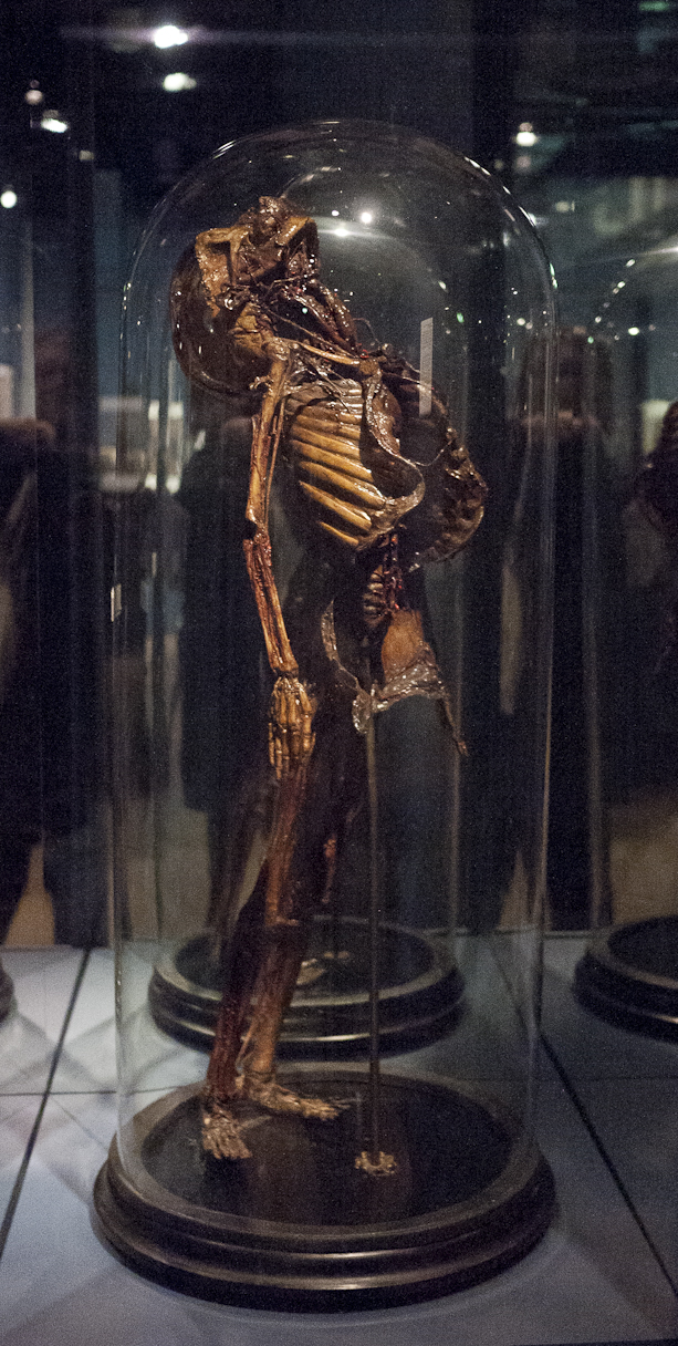
After a short stint at the Museum of London's 'Doctors, Dissection and Resurrection Men' this year I’m happy to say the above specimen – a corrosion cast of a child covered in shellac - has found its new home here at Barts Pathology Museum. It was originally housed at our sister hospital The Royal London, which is of course the home of Joseph Merrick (The Elephant Man) and in fact it was Frederick Treves, the man who ‘discovered’ the Elephant Man, who actually purchased this specimen on a trip to Paris in the 1800s.
We don’t have much more information about the child except that it probably pre-dates Treves by a long way and is from the early 1800s.

‘Corrosion casting’ similar to this was developed by such pioneers as Jan Swammerdam (above, Wikicommons) and Frederick Ruysch (below, Wikicommons) as early as the 16th century and was still popular with eighteenth- and nineteenth-century anatomists. Swammerdam and Ruysch had created a technique to inject the blood and lymphatic vessels of corpses with a mixture of wax, talc and pigments that set and endured, and often the surrounding flesh was corroded or removed. Anatomists were still using Swammerdam’s syringe to inject mixtures of varying proportions into their specimens until the early 20th Century.

According to Museum Vrolik, Ruysch had created the art of injecting specimens with a wax-like substance, dyed red with cinnabar, so that they could be more lifelike when displayed in jars (similar to the pink-tinted embalming fluid used to make loved ones more lifelike today.) However, he also injected tissues and organs which he then ‘embalmed and dried’ which would be more likely to give the dehydrated look of this specimen. In addition to such methods Ruysch introduced a new way of embalming specimens (based upon techniques already known by the ancient Egyptians).”

The passage reads: “Examples of organs in the Vrolik collection that have been injected with wax or dried include placentas and penises and, in particular, human and animal hearts. In order to make their anatomical structure more visible, in some cases one half of the heart was injected with red wax and the other half of the organ was injected with dark blue wax. Dry specimens, like bone and certain membranes, were dehydrated by exposing them to the external environment and subsequently coating them with a kind of varnish. The majority of the dry organ and tissue specimens were coated with shellac after drying.” A fate which seems to have befallen our shellac child in his entirety.
Shellac is an incredibly versatile substance which is secreted by the female Laccifer lacca (Lac Beetle), an insect of the order Hemoptera or the ‘true bugs’. It’s well known for it uses as a varnish for garden fences, paintings and musical instruments, but perhaps more surprising is the prevalence of edible shellac compounds in the pharmaceutical and food industries: it is found on medicinal capsules, in the wax of apples and lemons and on confectionery such as M&Ms. Used to limit water loss and prevent dessication, as well as to limit entry of pathogens, it is fairly obvious why this would be a good preservative for prepared anatomical specimens.

The interesting thing about this specimen is that it looks old and of its time – perhaps by using techniques mirroring those of the Ancient Egyptians, Ruysch ensured that an appearance of antiquity was an inevitable by-product. Medical students faced with a specimen such as this today possibly would not value its use as a teaching aid as much as they would have in those heady days of the 18th and 19th Centuries and would relegate it to the realms of ‘interesting’ and ‘unusual’ artwork but nothing more.
However that shouldn’t be the case – a study from 2011 was carried out to illustrate the use of shellac as a modern preservation method to replace the dangerous and carcinogenic formalin/formaldehyde which is currently used in dissection rooms. This study (Ref 2) has shown that shellac will preserve a new cadaver indefinitely in a way that is non-toxic, and said cadaver can also be placed into a softening room and in three days’ time be ready for student to use in their dissections. So it seems that an outdated yet beautiful specimen such as this can be used to inspire future generations of medical students and will also be a fascinating talking point on the ground floor of Barts Pathology Museum.
You can find out more about Barts Pathology Museum by clicking here. To can check out their new blog by clicking here. You can find out more about Carla Valentine by clicking here.
Image credits: Top image: Joanna Ebenstein; all other images of the specimen: Barts Pathology Museum, QMUL; portraits: wikimedia
Image credits: Top image: Joanna Ebenstein; all other images of the specimen: Barts Pathology Museum, QMUL; portraits: wikimedia
1 comment:
How brave those scientists were! Those days any kind of anatomical studies were frowned upon. Great blog.
Post a Comment